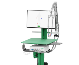
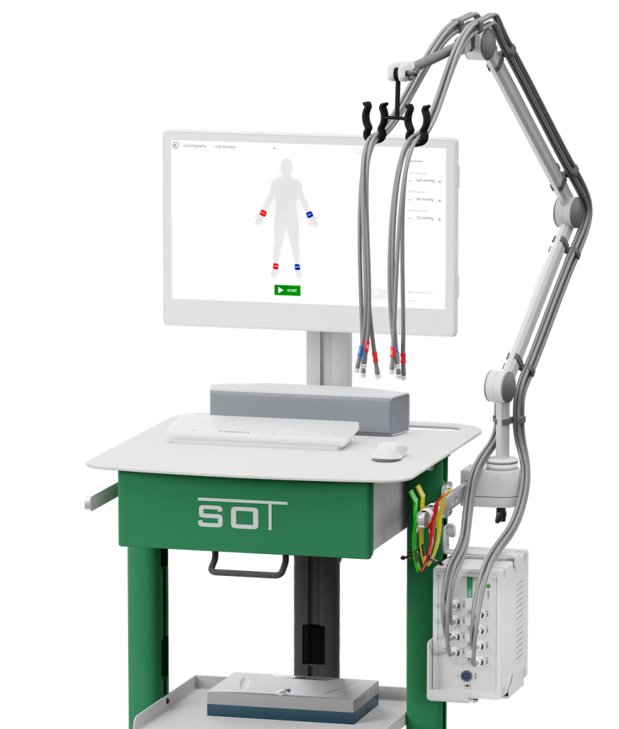
AngE™ COMPLETE
Fully Equipped Vascular Diagnostics System
🇦🇹 Made in Austria
AngE Solutions Brochure – 1.89 MB
Request now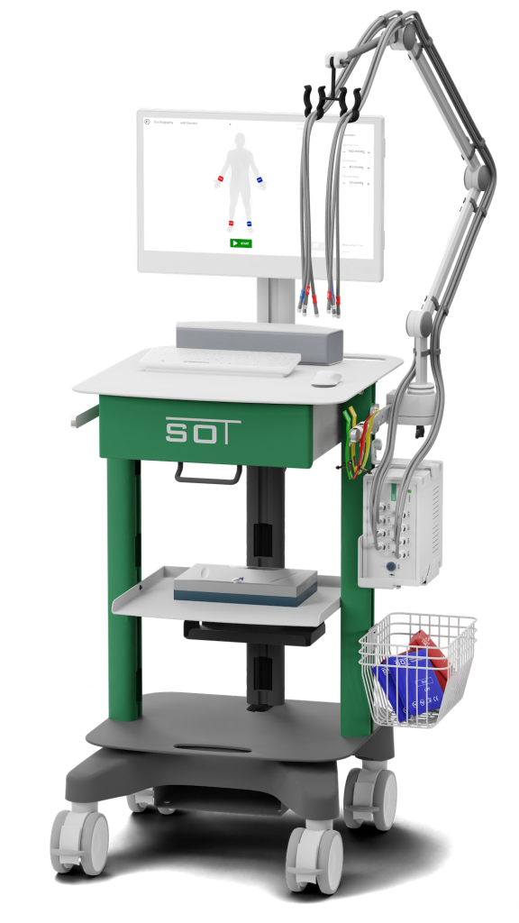
Test Protocols
Included or extendable measurement methods.
Segmental 8-Channel
Swiss Method
Segmental Pressure
SBP – Segmental Blood Pressures

Doppler
Bidirectional 4 and 8 MHz
Vascular Screening
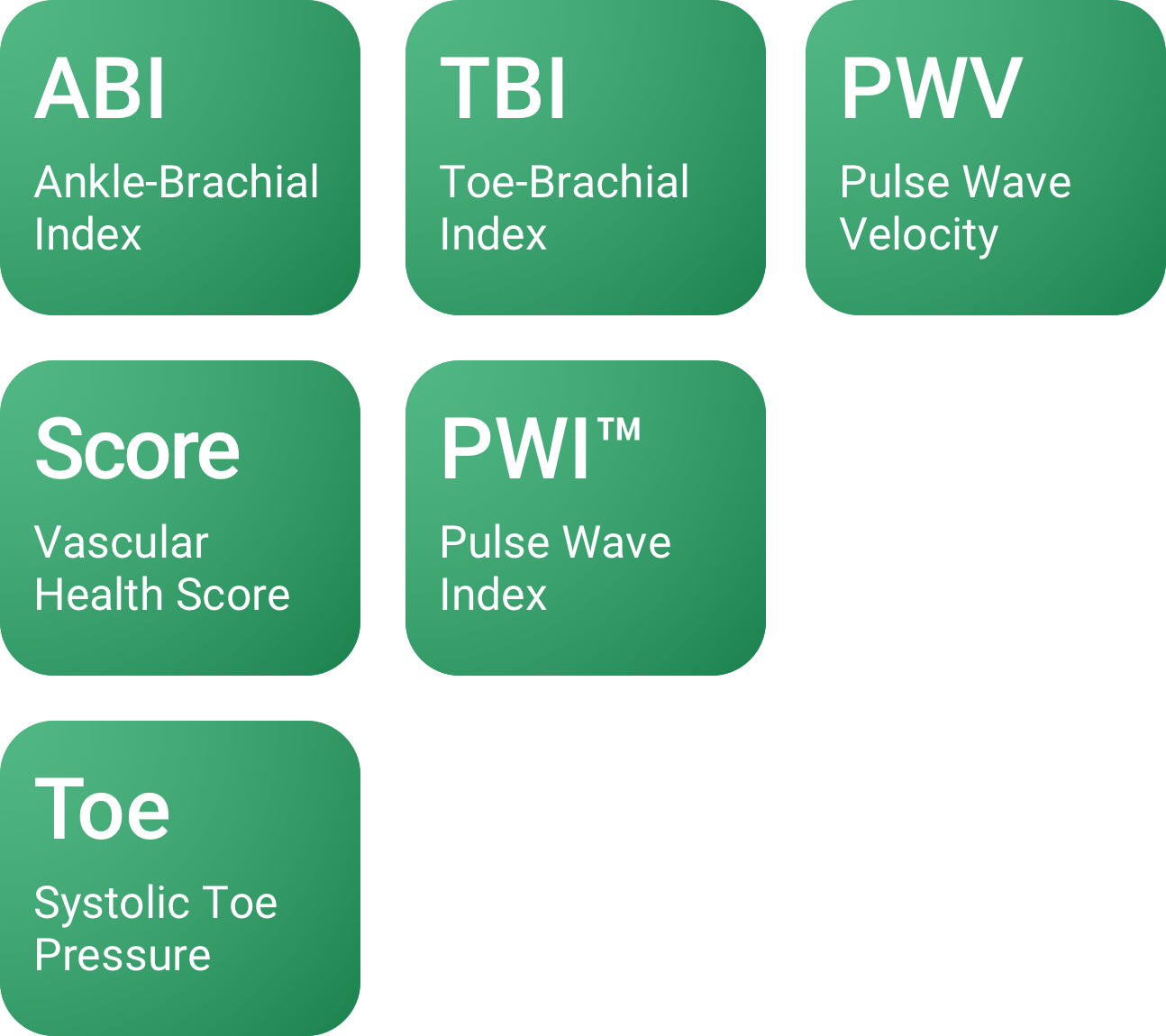
ABI, TBI, PWI™ and Score
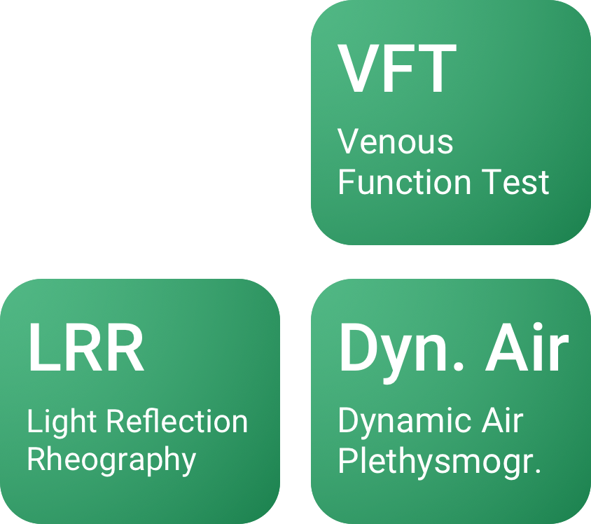
VOP
Venous Occlusion Plethysmogr.
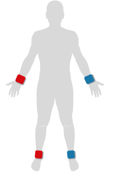
ABI
Ankle Brachial Index Test
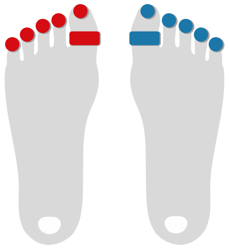
TBI
Toe Brachial Index
CVI
Chronic Venous Insufficiency
Toe Pressure
Systolic Toe Pressure

Erectile Dysfunction
PBI – Penile Brachial Index
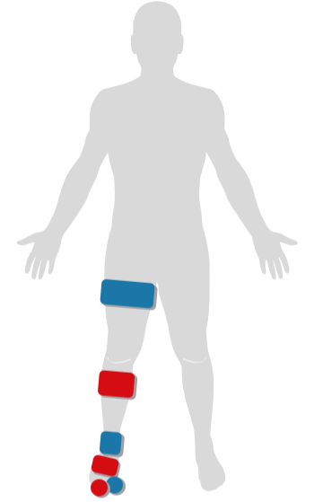
Segmental
Segmental Oscillography
Raynaud's Syndrome
Morbus Raynaud Test

TOS
Thoracic Outlet Syndrome
Stress Test
Exercise ABI Screening

Allen's Test
Palmar Arch Test

Finger Flow
Microcirculation
Toe Flow
Microcirculation
Add Custom Tests
Protocols and Report Templates
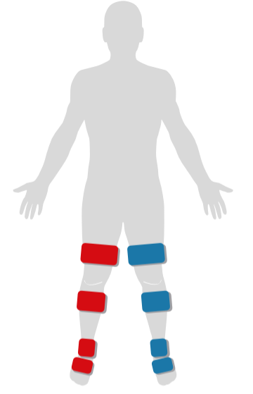
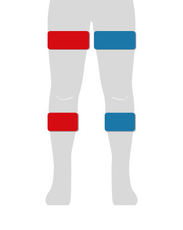
8-Channel Segmental Oscillography
Perform a simultaneous measurement on up to 8 cuff positions simultaneously to efficiently localize the level of an occlusion.
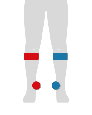
Venous Occlusion Plethysmography (VOP)
Assess the venous status of the legs in lying position by performing plethysmographic measurements, such as Reactive Hyperaemia Test and Dynamic Venous Air Plethysmography, with cuffs only.
PWI™ – Pulse Wave Index
The Pulse Wave Index is a clear and sensitive parameter based on pulse wave shape to determine the severity of blood flow disorders in significantly less time.
Micro- and Macrocirculation
Assess the Micro- and Macrocirculation to estimate the Wound Healing success.
Stress Tests
Record the patients' blood circulation after passive and active stress tests, such as knee bends and toe tip stands, for effective clarification.
Venous Function Measurements
AngE Phlebo calculates pump volume (V0), venous fill time (T0) and venous half-life time (T50) automatically. The results are stated in a simple traffic light display and allow for a fast venous valve incompetence diagnostics.
Arterial Blood Flow Tests
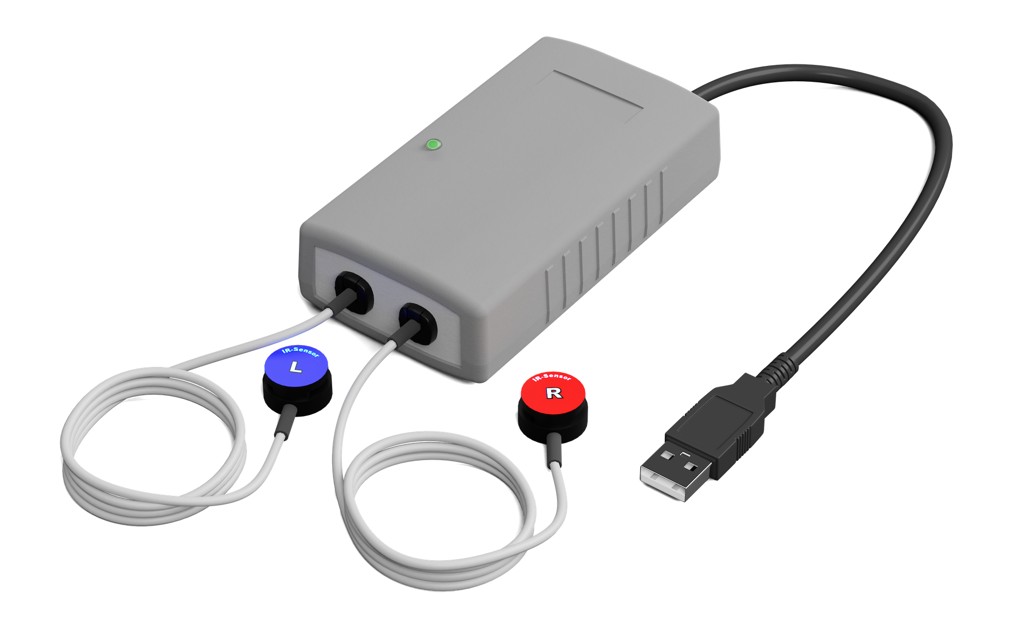
By applying the optical sensors on toes or fingers, arterial circulation disorders such as Morbus Raynaud or TOS can easily be assessed.
Pulse Wave Velocity and HRV
Determine Pulse Wave Velocity (PWV) and Heart Rate Variability (HRV) by recording the ECG-Trigger.
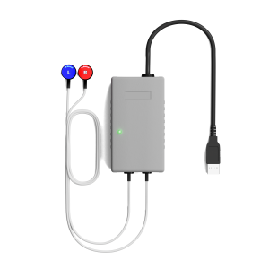
Doppler ABI
Use the 8 MHz or 4 MHz doppler probes to assess and record the blood flow vessel by vessel.
Detailed Report Printout
The one-page report combines all important details about the patient’s vascular status on a single page at the touch of a button.
Comprehensive Software
The AngE-System comes with a sophisticated macOS and Windows software featuring patient management, measurement analysis, DICOM/HL7 interfaces and many more.
Flexibility
A modular design ensures a tailor-made and future-proof investment.
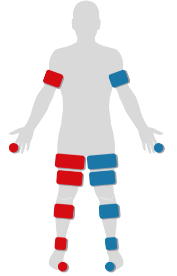
8-Channel Segmental Oscillography
The simultaneous recording of up to eight channels allows for the localization of occlusions as well as the automated calculation of pulse wave parameters within 45 seconds.
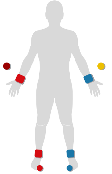
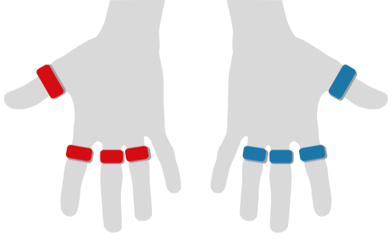
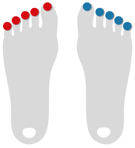
Finger & Toe Pressure
The OPO module allows performing optical pulse oscillography to determine blood flow in fingers and toes as well as to conduct arterial pressure measurements.
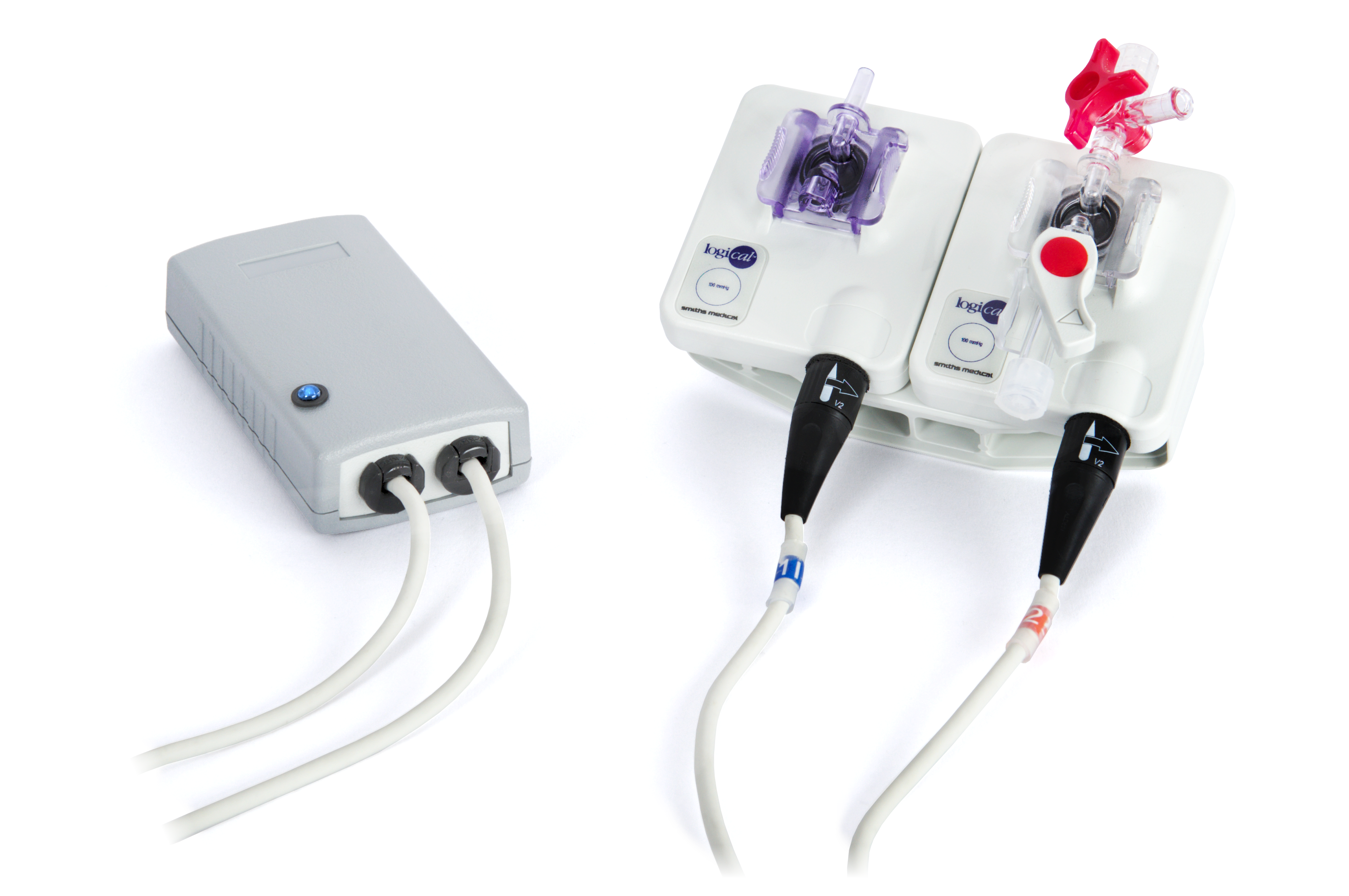
Ultrasonic Doppler
The AngE combines ultrasonic probes with pneumatic cuffs to allow Doppler pressure measurements with up to 16 tracks. The Doppler indices can be displayed at a glance on a dedicated overview report.
Venous Air Plethysmography
The Air VVP allows conducting plethysmographic measurements by using cuffs only. The often used mercury-filled strain gauges become unnecessary.
AngE Phlebo
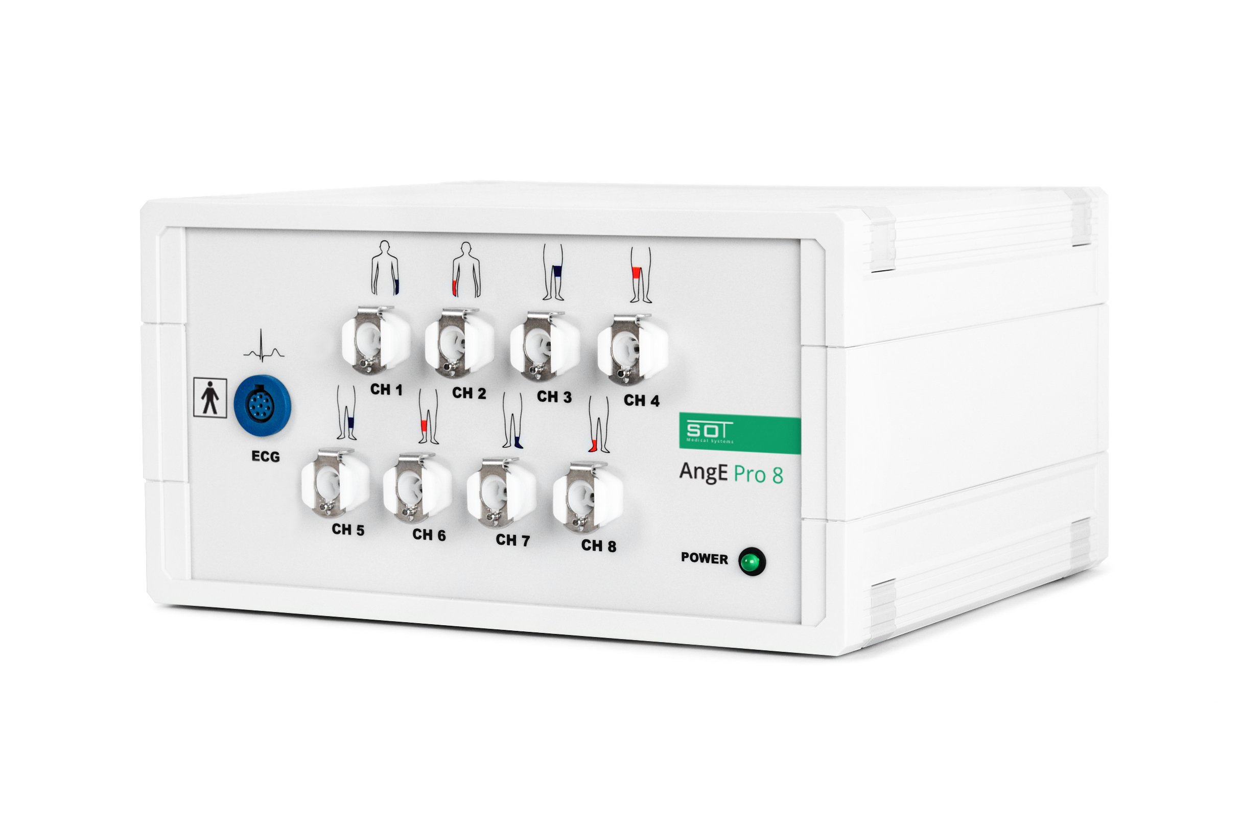
AngE Pro 8
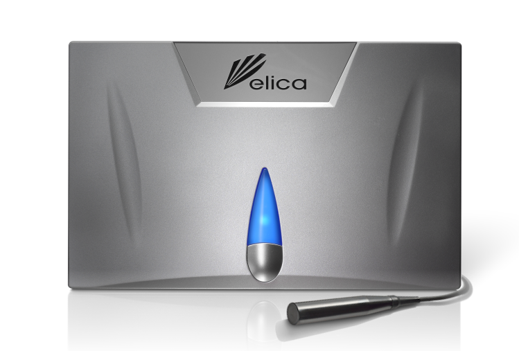
AngE Doppler
AngE PDM
Main Measurement-Parameters
| oABI (oscillometric Ankle-Brachial-Index) | TBI (Toe-Brachial-Index) | PWI™ (Pulse-Wave-Index) | HRV (Heart-Rate-Variability) |
| Central PWV (Pulsewave Velocity) | Toe-Pressure / Finger-Pressure | Skin-Temperature | V0 (Venous Pump Volume) |
| T0 (Venous Fill-Time) | T50 (Venous Half-Life Time) | Doppler-ABI | RI (Reflexion Index) |
| SI (Stiffness Index) | Venous Peak-Flow | Arterial In-Flow | Invasive Compartment-Pressure |
Recent Scientific Publications
"Time consumption for testing was significantly lower using the semiautomated AngE-device – a finding that is economically important, especially in high volume centers and epidemiologic studies."
– Mayr, Hirschl, Klein-Weigel, Girardi, Kundi; 2019
"Tissue optical perfusion pressure: a simplified, more reliable, and faster assessment of pedal microcirculation in peripheral artery disease."
– Horstick, Messner, Grundmann, Yalcin, Weisser, Espinola-Klein; 2020
The high sensitivity of the optical sensors allow for a good documentation of the pulse waves, even with marginal blood flow. Given the virtually unfiltered display of pulse curves, dicrotic waves can be clearly identified for healthy and elastic arteries.

The semi-automated vascular screening with the AngE™ AngioExperience is after more than 150 years a long overdue replacement of simple doctors blood pressure recordings. By simultaneous recordings of pressure, pulse and ECG signals and their mathematical computations individual vascular functions can be assessed beyond an epidemiological cardiovascular risk assessment.

A highly effective, easy-to-use system that performed exceptionally well in practical testing and is particularly useful in general practice, especially when determining further treatment options and differential diagnoses.

The AngE Phlebo is the state-of-art, haemodynamically significant D-PPG system for venous diagnostics. This non-invasive functional investigation has always helped me accurately examine venous disorders, even with complex cases.
We have been using the AngE ABI+ system for several weeks now and are extremely satisfied. The examination is quick and straightforward, integrates seamlessly into our workflow, and provides a wide range of important vascular parameters in a clear and patient-friendly report. We highly recommend it for preventive medicine and cardiology applications.

Because it is easy to use and highly accurate, this method is ideal for screening; the standard measurements can be easily delegated to medical assistants. I therefore prefer this system over other similar devices currently available on the market.
The ease of use and the clear visualization of arterial and venous vascular function make the AngE an indispensable part of our daily practice. As Vienna’s largest wound care center, we particularly benefit from the support it provides in wound care and in the diagnosis of lipedema and lymphedema.

Request Information
Submit the form to be contacted by one of our AngE COMPLETE Experts.
References
1. Automated oscillometric blood pressure and pulse-wave acquisition for evaluation of vascular stiffness in atherosclerosis. (Massmann et al. 2017)
2. Diagnostic Accuracy Study of an Oscillometric Ankle-Brachial Index in Peripheral Arterial Disease: The Influence of Oscillometric Errors and Calcified Legs. (Herráiz-Adilo et al. 2016)
3. Interrater and intrarater reliability of photoplethysmography for measuring toe blood pressure and toe- brachial index in people with diabetes mellitus. (Scanlon C. et al. 2012)
4. Photoplethysmography detection of lower limb peripheral arterial occlusive disease: a comparison of pulse timing, amplitude and shape characteristics. (Allen J et al. 2005)
5. Venous filling time using air-plethysmography correlates highly with great saphenous vein reflux time using duplex. (Lattimer et al. 2014)


38 Years of
Vascular Experience
With more than 4,000 installations in more than 16 countries, SOT secures its position as knowledge leader in vascular diagnostics. We have made our knowledge available on:

Instant Support
in one minute
Our product experts in Austria will help you with remote support without any waiting time or ticket system. Call us during office hours and we will help you within a minute.
+43 4227 84 991
Mo - Th: 8am - 5pm, Fr: 8am - 12am
Made in
Austria 🇦🇹
We develop and produce our AngE™ products in Austria, where we adhere to the highest quality standards (ISO 13485:2016) and invest in our leading vascular diagnostic systems.