Venous diseases are among the most common vascular disorders worldwide, affecting millions of people and significantly impacting quality of life. Symptoms such as heavy legs, swelling, varicose veins, skin changes, and venous ulcers often result from impaired venous return caused by chronic venous insufficiency (CVI).
Early assessment of venous function plays a key role in identifying venous reflux before symptoms worsen. While duplex ultrasound remains the gold standard for anatomical evaluation, photoplethysmography (PPG) offers a fast, non-invasive, and cost-effective method for assessing venous function in everyday clinical practice.
This guide explains how to perform a venous function test using PPG, how to ensure reliable measurements, and how to interpret the results.
What Is a Venous Function Test?
A venous function test evaluates how effectively blood returns from the lower limbs to the heart.
Unlike duplex ultrasound, which visualizes veins and blood flow directly, PPG assesses venous function by measuring changes in blood volume within the superficial microcirculation.
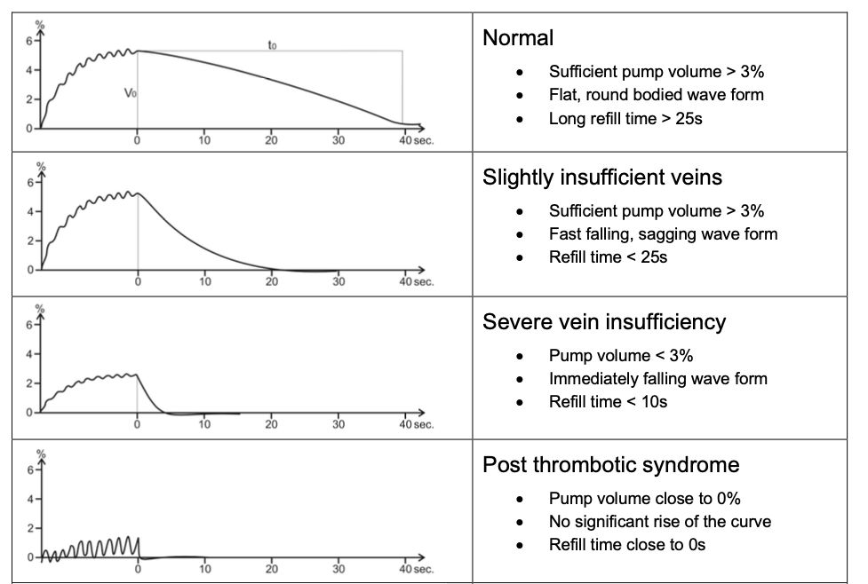
After activating the calf muscle pump through repeated foot movements, the system measures how quickly blood refills the superficial veins. This parameter is known as the Venous Refill Time (VRT).
A shortened VRT may indicate venous reflux caused by incompetent venous valves.
When Should a Venous Function Test Be Performed?
Venous function testing is recommended for patients presenting with symptoms or risk factors associated with chronic venous disease, including:
- Heavy or tired legs
- Leg swelling (edema)
- Varicose veins
- Skin discoloration or venous eczema
- Venous leg ulcers
- Suspected chronic venous insufficiency
- Follow-up after conservative or surgical treatment
As a rapid screening method, PPG can help determine whether further diagnostic evaluation is required.

Equipment Required
Before beginning the examination, ensure that the following equipment is available:
- PPG sensor
- Venous function measurement system
- Examination chair
- Alcohol wipe (if necessary)
- Clean, dry skin
The patient should be seated comfortably with both feet flat on the floor throughout the measurement.
Step 1 – Prepare the Patient
Before starting the examination, explain the procedure to the patient..
The patient should sit comfortably with the hips and knees forming an angle of approximately 110°, allowing the lower legs to be slightly extended forward. Both feet should rest flat on the floor, and shoes and socks should be removed to expose the lower legs.
The skin should be clean and dry to ensure optimal sensor contact.
Standardized patient positioning is essential for obtaining reliable and reproducible measurements.
Step 2 – Position the PPG Sensor
Place the PPG sensor approximately 10 cm above the medial malleolus (inner ankle) on the lower leg.
Correct sensor placement is essential because inaccurate positioning may affect the quality of the recorded signal and the calculated Venous Refill Time.
The sensor should adhere firmly to the skin without excessive pressure.
Step 3 – Activate the Calf Muscle Pump
The patient performs approximately 10 dorsal flexion movements of the foot to activate the calf muscle pump. These rhythmic movements empty the superficial veins and reduce venous pressure in the lower leg.
Once the movements are completed, the patient remains completely still while venous refilling is recorded automatically.
Step 4 – Measure the Venous Refill Time
Once the exercise has finished, the PPG device continuously records changes in blood volume as the veins refill.
The software automatically calculates the Venous Refill Time (VRT).
No additional patient movement is required during this phase.
The entire examination typically takes only a few minutes.
Understanding the Results
Venous Refill Time reflects how efficiently the venous valves prevent blood from flowing backward after muscle contraction.
In healthy veins, refill occurs gradually because competent valves maintain one-way blood flow toward the heart.
If venous valves are insufficient, blood refluxes into the superficial veins, causing a significantly shorter refill time.
Although normal reference values may vary slightly depending on the system and protocol used, a shortened VRT generally indicates impaired venous function and warrants further clinical evaluation.
Results should always be interpreted together with the patient’s symptoms, medical history, and, when appropriate, duplex ultrasound findings.
How to Differentiate Between Superficial and Deep Venous Insufficiency
A standard PPG examination provides information about the overall function of the venous system by measuring the Venous Refill Time (VRT). However, it does not distinguish whether venous reflux originates from the superficial or the deep venous system.
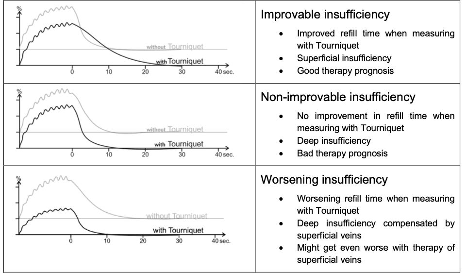
To differentiate between the two, the examination can be repeated using a tourniquet cuff placed above or below the knee. The cuff temporarily compresses the superficial veins while leaving the deep venous system unaffected.
The results can then be interpreted as follows:
- Improved refill time with the tourniquet applied indicates that the superficial veins are the primary source of reflux, suggesting superficial venous insufficiency.
- No improvement in refill time suggests that reflux originates from the deep venous system, indicating deep venous insufficiency.
- A worsening refill time may indicate that the superficial veins are compensating for impaired deep venous function. In these cases, treatment decisions should be made carefully, as eliminating superficial veins could further impair venous drainage.
This simple additional measurement provides valuable functional information and supports treatment planning before further diagnostic or therapeutic interventions.
Common Mistakes During Testing
Reliable results depend on standardized examination procedures.
Common sources of measurement error include:
- Incorrect sensor placement
- Poor skin contact
- Patient movement during measurement
- Incomplete foot exercises
- Failure to remain still after calf muscle activation
Following a standardized protocol helps ensure reproducible and clinically meaningful results.
Why Use PPG for Venous Screening?
Photoplethysmography offers several advantages for routine vascular assessment:
- Fast examination time
- Non-invasive and painless procedure
- Objective assessment of venous function
- Easy integration into daily clinical workflow
- Suitable for screening and follow-up examinations
- Supports early detection of chronic venous insufficiency
Although duplex ultrasound remains indispensable for anatomical assessment, PPG provides valuable functional information that complements comprehensive venous diagnostics.
Modern Venous Function Assessment
Today’s vascular diagnostic systems combine automated measurement protocols with digital documentation, allowing clinicians to perform standardized venous function testing quickly and efficiently.
Solutions such as AngE™ ABI+ integrate photoplethysmography into a standardized vascular assessment workflow, enabling efficient and reproducible venous function testing alongside additional vascular diagnostic parameters.
Conclusion
Venous function testing using photoplethysmography is a simple, fast, and reliable method for assessing venous insufficiency.
By following a standardized testing protocol—including proper patient preparation, accurate sensor placement, correct calf muscle activation, and careful interpretation of Venous Refill Time—clinicians can obtain reproducible results that support early diagnosis and ongoing patient management.
As part of a comprehensive vascular assessment, PPG provides valuable functional insights that complement clinical examination and duplex ultrasound, helping improve the detection and monitoring of chronic venous disease.
Sources:
https://vascular-academy.com/venous-tests/d-ppg-muscle-pump-test-venous-reflux/