Thoracic Outlet Syndrome (TOS) is a complex condition caused by compression of nerves or blood vessels in the anatomical passage between the neck and shoulder. Because symptoms can be intermittent, position-dependent and similar to those of other musculoskeletal or neurological disorders, TOS can be difficult to recognize.
When vascular involvement is suspected, functional and positional vascular testing can provide valuable objective information about changes in blood flow during provocative maneuvers.
What Is Thoracic Outlet Syndrome?
The thoracic outlet is the narrow anatomical space between the lower neck and the armpit through which the brachial plexus, subclavian artery and subclavian vein pass toward the upper extremity.
Compression of these structures can result in three main forms of TOS:
- Neurogenic TOS – compression or irritation of the brachial plexus
- Venous TOS – compression of the subclavian vein
- Arterial TOS – compression of the subclavian artery
Neurogenic TOS is the most common form, while venous and arterial TOS are less frequent but can have important vascular consequences.
The condition can be associated with anatomical factors, previous trauma, repetitive strain or activities that repeatedly place stress on the shoulder and upper extremity.
When Should You Suspect TOS?
The first step in screening is recognizing symptoms that may indicate neurovascular compression.
Depending on the structures involved, patients may report:
- Pain in the neck, shoulder or arm
- Numbness or tingling in the hand or arm
- Arm weakness or fatigue
- Swelling of the arm or hand
- Coldness or discoloration of the hand
- A feeling of heaviness in the arm
- Symptoms that occur or worsen when the arm is elevated or placed in a specific position
A particularly important clue is position-dependent symptoms. A patient may be relatively asymptomatic at rest but develop symptoms during arm elevation, shoulder depression or other specific movements.
This dynamic nature is one reason why a conventional examination at rest may not fully reveal the underlying problem.
Why Is TOS Difficult to Diagnose?
TOS can mimic several other conditions, including cervical spine disorders, peripheral nerve compression and musculoskeletal problems.
In addition, vascular compression may only occur when the patient adopts a particular posture. This means that blood flow can appear relatively normal in a resting position while becoming impaired during provocation.
There is therefore no single test that can independently diagnose TOS. Clinical assessment generally combines medical history, physical examination, provocative testing and, when indicated, vascular or imaging studies.
How to Screen for Vascular TOS
When arterial or venous involvement is suspected, the objective is to determine whether a specific position produces measurable vascular changes.
A practical vascular screening approach can be divided into four steps:
Step 1 – Clinical Assessment
Begin with a detailed history and physical examination.
Assess:
- The patient’s symptoms and their distribution
- Whether symptoms are unilateral or bilateral
- Activities or positions that trigger symptoms
- Previous trauma or repetitive upper-extremity activity
- Changes in skin color or temperature
- Swelling or venous congestion
- Peripheral pulses and differences between the arms
The relationship between symptoms and arm position is particularly relevant when considering TOS.
Step 2 – Perform Provocation Testing
Because TOS is often a dynamic, position-dependent disorder, provocative maneuvers are used to reproduce the conditions under which compression may occur.
Depending on the protocol, maneuvers may include:
Head rotation and deep inspiration
The patient turns the head toward the affected side, elevates the chin and takes a deep breath. An additional downward pull of the arm may be applied.
A reduction or disappearance of the radial pulse and reproduction of symptoms may occur during this maneuver.
Arm elevation
The patient raises the arm above shoulder level.
Compression of the neurovascular structures may become more pronounced in this position, potentially resulting in changes in the arterial pulse waveform.
Shoulder depression
Lowering or pulling the shoulder downward can also narrow the thoracic outlet and may reproduce symptoms or vascular changes.
Important: Provocative maneuvers can support the clinical assessment, but a change in pulse alone is not sufficient to establish a diagnosis. The Society for Vascular Surgery specifically notes that loss of the radial pulse during an Adson maneuver can also occur in people without TOS.
Step 3 – Measure Blood Flow Changes Objectively
Instead of relying exclusively on palpation of the radial pulse, modern vascular screening can continuously record pulse waveforms during the provocative maneuvers.
For this purpose, optical photoplethysmography (PPG) sensors can be placed on the index fingers of both hands.
The sensors detect changes in peripheral blood volume and generate pulse waveforms that can be compared between the two sides while the patient moves through the different test positions.
This makes it possible to observe whether a provocative position causes:
- A reduction in pulse-wave amplitude
- Changes in pulse-wave morphology
- Delayed pulse-wave rise
- Differences between the left and right side
- A substantial reduction or disappearance of pulse oscillations
A significant reduction or disappearance of pulse oscillations during a provocative maneuver may indicate vascular compression and can support further evaluation for arterial TOS.
Step 4 – Evaluate the Measurement Parameters
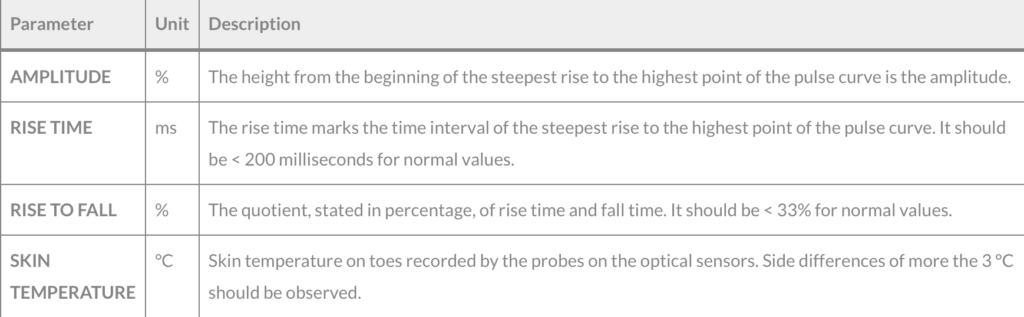
A PPG-based TOS assessment can provide several parameters for evaluating the pulse waveform.
What Does a Positive TOS Screening Test Mean?
A positive or suspicious vascular screening result does not automatically mean that the patient has TOS.
Instead, the test can indicate that a particular arm position is associated with measurable changes in peripheral blood flow.
This information can help answer an important clinical question:
Does the patient’s vascular function change when the thoracic outlet is placed under provocation?
If significant changes are observed, further diagnostic evaluation may be appropriate.
For vascular TOS, this may include duplex ultrasound with provocative maneuvers. Depending on the clinical situation, CTA or MRA can provide additional anatomical information and help demonstrate vascular entrapment.
Screening vs. Diagnosis: Why the Difference Matters
TOS should be approached as a multimodal diagnostic challenge.
A functional screening test can identify changes in blood flow during provocation, but it does not replace clinical assessment or confirm the underlying anatomical cause.
A useful clinical pathway is therefore:
Symptoms → Clinical assessment → Provocation testing → Functional vascular assessment → Further vascular/imaging evaluation if indicated
This approach is particularly relevant because anatomical compression alone does not necessarily prove clinically significant TOS. The findings need to be correlated with the patient’s symptoms and clinical presentation.
Why Functional Screening Can Add Value
Traditional imaging provides important anatomical information. However, TOS is not purely an anatomical condition.
The degree of compression can change depending on:
- Arm position
- Shoulder position
- Muscle activation
- Posture
- Individual anatomy
This makes dynamic assessment particularly relevant. Recent clinical perspectives describe TOS as a position-provoked disorder in which findings that appear normal at rest may become abnormal when the arm is elevated or the shoulder is loaded.
Functional vascular testing can therefore complement anatomical imaging by showing what happens to blood flow during provocation.
A Practical TOS Screening Workflow
For clinical practice, the process can be summarized as follows:
1. Identify suspicious symptoms
Position-dependent pain, numbness, weakness, swelling, discoloration or coldness.
↓
2. Take a detailed history and perform a physical examination
Assess symptoms, pulses, skin changes and provoking activities.
↓
3. Perform standardized provocative maneuvers
Evaluate the patient’s response to specific arm, shoulder and head positions.
↓
4. Record vascular changes objectively
Use PPG or another appropriate vascular measurement method to monitor pulse-wave changes.
↓
5. Compare the findings
Evaluate baseline versus provocation and left versus right side.
↓
6. Refer for further diagnostic evaluation when indicated
Duplex ultrasound and, depending on the clinical question, CTA, MRA or other specialized testing may be required.
For objective, non-invasive vascular screening, the TOS test can be performed with the AngE™ Phlebo or AngE™ ABI+ systems. Both systems enable PPG-based pulse waveform analysis during provocative maneuvers, supporting the assessment of position-dependent changes in peripheral blood flow.
Conclusion
Thoracic Outlet Syndrome can be difficult to recognize because symptoms are often non-specific and may only appear when the upper extremity is placed in a particular position.
A structured screening approach can help identify patients in whom vascular compression should be investigated further. Provocation testing combined with objective pulse-wave measurement provides a way to evaluate dynamic changes in peripheral blood flow rather than relying solely on symptoms or pulse palpation.
For suspected vascular TOS, functional screening should be viewed as one component of a broader diagnostic pathway, supporting clinical assessment and helping determine when further vascular imaging or specialist evaluation is warranted
This article is intended for healthcare professionals and educational purposes. TOS diagnosis should be based on the complete clinical picture and appropriate specialist assessment.
Sources:
SOT- Blog: Thoracic Outlet Syndrome