The Ankle-Brachial Index (ABI) is an established, non-invasive parameter used to assess arterial perfusion of the lower extremities and support the detection of Peripheral Artery Disease (PAD).
Traditionally, ABI is measured manually using blood pressure cuffs and Doppler ultrasound. The modern oscillometric approach uses an automated system to record pressure oscillations at multiple extremities simultaneously. This provides a standardized and efficient measurement and enables direct comparison between the right and left sides.
In this guide, we explain how an automated ABI measurement is performed step by step and how the results can be interpreted.
The traditional ABI compares ankle and brachial systolic pressures.
The oscillometric ABI uses a different measurement principle. During cuff deflation, the system detects pressure oscillations generated by the pulsating arteries underneath the cuff. The maximum oscillation amplitude corresponds to the Mean Arterial Pressure (MAP).
For the SOT oscillometric approach, the oABI is calculated from the relationship between the mean arterial pressure at the ankle and the mean arterial pressure at the upper extremity/wrist. The Vascular Academy describes this parameter as the Oscillometric Ankle-Wrist ABI (oABI).
The principle:
oABI = Mean Arterial Pressure at the ankle ÷ Mean Arterial Pressure at the arm/wrist
This differs from the conventional Doppler ABI, which is based on systolic pressures.
Why Use an Automated ABI?
The traditional ABI measurement requires the clinician to measure the extremities individually using a cuff and Doppler.
The oscillometric approach uses simultaneous measurements, allowing the system to record the pressure oscillations of the extremities during the same examination. The Vascular Academy describes the method as operator-independent and time-efficient, while also highlighting the possibility of directly comparing pulse-wave signals from both sides.
An automated approach can therefore provide:
- Standardized measurement
- Simultaneous assessment
- Reduced operator dependency
- Direct left-right comparison
- Automated calculation
- Digital documentation
How to Perform an Automated ABI
The measurement can be performed in just a few simple steps.
Step 1 – Prepare the Patient
The patient should be positioned comfortably and remain relaxed throughout the examination.
The arms and legs should be supported and positioned comfortably. The patient should not move or speak during the measurement, as movement can introduce artefacts into the oscillometric signals.
Before starting, ensure that clothing does not interfere with cuff placement.
Step 2 – Position the Cuffs
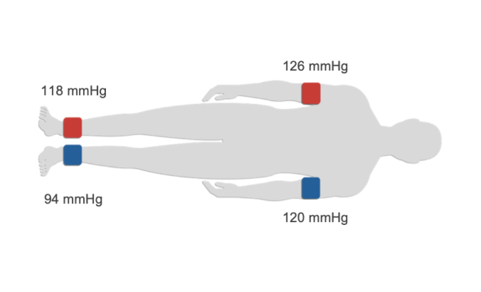
The automated ABI measurement uses four cuffs to simultaneously assess both sides.
The cuffs are positioned on:
Right upper extremity
Left upper extremity
Right ankle
Left ankle
Depending on the measurement setup, the upper-extremity cuffs can also be positioned at the wrists.
This configuration allows the system to record the oscillations from all measurement sites simultaneously. The Vascular Academy specifically describes the use of cuffs at the ankles and upper arms, with the wrist as an alternative position.
Step 3 – Start the Automated Measurement
Once the cuffs are positioned correctly, start the automated measurement.
The system inflates and deflates the cuffs according to the defined measurement protocol.
As the cuff pressure changes, the system records the oscillations caused by arterial pulsation.
The maximum oscillation amplitude is used to determine the mean arterial pressure at each measurement site. These pressure signals are then used to calculate the oscillometric ABI.
The patient should remain still and avoid talking until the measurement is complete.
Step 4 – Review the Results
After the measurement, the system automatically displays the results for both sides.
The clinician can review:
- Mean arterial pressure
- oABI
- Pulse-wave amplitude and morphology
- Side-to-side differences
- Additional parameters depending on the system configuration
The simultaneous measurement makes it possible to compare the right and left extremities directly. Differences in pressure or pulse-wave characteristics may provide additional information about arterial circulation.
Watch the Measurement in Practice
The video demonstrates the automated four-cuff ABI measurement with AngE™ ABI+, including the measurement setup and automated workflow.
How to Interpret the ABI
For clinical interpretation, ABI categories should be considered together with the patient’s symptoms, medical history and other vascular findings.
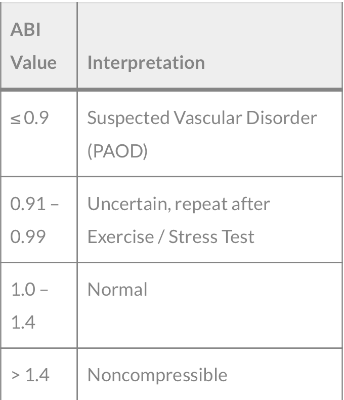
An ABI of ≤ 0.90 is considered abnormal and supports the diagnosis of PAD in the appropriate clinical context. Values above 1.40 indicate noncompressible arteries and may require additional physiological assessment.
The SOT oscillometric ABI (oABI) is based on MAP-derived oscillometric measurements and should be interpreted according to the measurement method and system used. The Vascular Academy describes oABI ≤ 0.9 as indicative of a vascular disorder/PAD.
What If the Result Is Inconclusive?
A single ABI value does not always provide a complete picture of arterial perfusion.
When arteries are noncompressible, the pressure measured at the ankle may be difficult to interpret. This can occur, for example, in the presence of medial arterial calcification.
According to the 2024 ACC/AHA guideline, a Toe-Brachial Index (TBI) with waveforms should be used when the resting ABI is >1.40.
Additional parameters such as Toe Pressure, TBI, pulse-wave analysis or exercise testing may therefore complement ABI measurement when clinically indicated.
Why Standardized Measurement Matters
Reliable vascular assessment depends on consistent measurement conditions.
An automated oscillometric approach can help standardize the examination by:
- Measuring multiple extremities simultaneously
- Reducing manual operator dependency
- Applying the same measurement protocol to both sides
- Automatically calculating the oABI
- Providing immediate side-to-side comparison
The Vascular Academy also emphasizes that the recorded pulse waves can be assessed in addition to the pressure-derived oABI, providing further information about arterial function.
Automated ABI with AngE™ ABI+
AngE™ ABI+ is designed for automated vascular screening using a four-cuff oscillometric measurement approach. The system can simultaneously assess the extremities and automatically calculate the ABI while providing additional pulse-wave information.
Depending on the configuration, the system can also be extended with additional vascular measurements, including TBI, Toe Pressure, PWI™, PWV and Venous Function Testing.
This allows the automated ABI to be integrated into a broader vascular screening workflow.
Conclusion
Automated oscillometric ABI measurement provides a fast, standardized and non-invasive approach to vascular assessment.
Instead of measuring each extremity individually, the four-cuff approach records pressure oscillations simultaneously and uses the resulting mean arterial pressure signals to calculate the oscillometric ABI.
With automated measurement, direct side-to-side comparison and digital documentation, oABI can be efficiently integrated into routine vascular screening.
When results are abnormal or inconclusive, additional parameters such as TBI and Toe Pressure can provide further information about peripheral arterial perfusion.
Sources
- SOT Medical Systems – Blog Post – Why ABI isn’t Enough: Why TBI and PWI Matter in Vascular Diagnostics
- Vascular Academy – ABI: Ankle-Brachial Index
- Vascular Academy – PVR / Pulse Volume Recording
- SOT Medical Systems – AngE™ ABI+